×

Cosmetic Surgery Blog
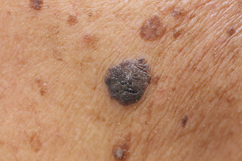
Melanomas usually just happen on their own!

The majority of melanomas arise from new lesions rather than existing moles, according to a review published online Aug. 29 in the Journal of the American Academy of Dermatology.

While we always emphasis the importance of following the appearance of your existing moles the fact is that most melanomas (70%) do not derive from existing moles “going bad” , but just develop as melanomas from the very start.

You should still follow your moles (30% of melanomas do derive from existing moles), but also know your whole skin so you can recognize any new moles. To this end we highly recommend full body digital photography as a baseline documentation of your moles and skin against which any potentially changing or new moles can be compared.

Researchers conducted a review of 38 previously published medical studies involving 20,126 melanomas.

The findings showed that only 29.1 percent of the skin cancers started in moles patients already had, while 70.9 percent arose as new lesions on the skin.

“In conclusion, in this systematic review and meta-analysis we found that less than one-third of melanomas were nevus-associated.”


Link to abstract:
http://www.jaad.org/article/S0190-9622(17)32051-0/abstract

Melanoma Rates Still Rising in California

A new study from the Journal of Investigative Dermatology has confirmed that the rates of melanoma are still on the rise among white Californians. All the more reason to protect yourself from the damaging rays of the sun, check your own skin routinely and have an annual skin cancer screening exam.

Title of article: “Continued increase in melanoma incidence across all socioeconomic status groups in California, 1998-2012”

Link to abstract:
http://www.jidonline.org/article/S0022-202X(17)31867-5/pdf

Thin Melanomas – Age Matters!

A new study suggests that in young patients with thin melanomas (0.5 -1mm depth) there may be an approximately 5% risk of lymph nodes being involved. As such, even though present guidelines do not recommend it, in young patients with melanomas between 0.5 and 1mm sentinel node biopsy may be considered.

Association Between Patient Age and Lymph Node Positivity in Thin Melanoma

Andrew J. Sinnamon, MD1; Madalyn G. Neuwirth, MD1; Pratyusha Yalamanchi, BA1; et alPhyllis Gimotty, PhD
JAMA Dermatol. Published online July 19, 2017. doi:10.1001/jamadermatol.2017.2497

Link to abstract:
http://jamanetwork.com/journals/jamadermatology/article-abstract/2643739

Mohs wins out again

Recent published study confirms Mohs micrographic surgery as the patients treatment of choice even for superficial skin cancers. Patients in the study preferred Mohs surgery not only because it gave a better long term cure rate but it was seen as a much more tolerable procedure compared to the alternative treatments.

Link to abstract:
https://insights.ovid.com/crossref?an=00042728-900000000-99021


Treatment Patterns, Outcomes, and Patient Satisfaction of Primary Epidermally Limited Nonmelanoma Skin Cancer

Dermatologic Surgery. Publish Ahead of Print():, JUN 2017
Benjamin A. Drew; Pritesh S. Karia; and 3 more

TAKE-HOME MESSAGE

    • In this retrospective cohort study, 550 patients with superficial basal cell carcinoma (BCC) and squamous cell carcinoma in situ (SCCis) were treated with either Mohs micrographic surgery (MMS) or non-MMS (5% 5-fluorouracil, cryotherapy, ingenol mebutate, or imiquimod). Although both groups had high 5-year recurrence-free survival, this was significantly higher with MMS compared with non-MMS (99% vs 95%, respectively; P = .004). Moreover, 97% of MMS patients were willing to undergo another treatment compared with 86% of non-MMS patients (P = .024). Results from a satisfaction survey offered to 25% of patients indicated that a prolonged treatment course and pain contributed to dissatisfaction on the part of the patients who did not undergo MMS.
    • This study found low recurrence rates with both surgical and nonsurgical treatments for superficial BCC and SCCis. However, MMS was associated with higher patient satisfaction and cure rates compared with non-MMS.

– InYoung Kim, MD, PhD

BACKGROUND

Epidermally limited nonmelanoma skin cancer (ELNMSC) (superficial basal cell carcinoma [SBCC] and squamous cell carcinoma in situ [SCCIS]) is common. Data on outcomes and patient satisfaction are lacking.

OBJECTIVE

To examine treatment efficacy and satisfaction in ELNMSC patients.

PATIENTS AND METHODS

Retrospective cohort study of adults with primary SBCC or SCCIS. A 25% random subset completed a satisfaction questionnaire.

RESULTS

Five hundred and fifty patients with 227 SBCC and 451 SCCIS were included; 329 tumors (49%) were treated with Mohs micrographic surgery (MMS) and 349 (51%) with non-MMS (imiquimod [n = 26], 5% 5-fluorouracil [n = 234], ingenol mebutate [n = 32], or cryotherapy [n = 57]). Five-year recurrence-free survival was high in both groups, with MMS having a small but statistically significant advantage (99% vs 95%, p = .004). More MMS patients were willing to undergo treatment again (97% vs 86%, p = .024). Dissatisfaction was mostly due to prolonged treatment course and pain associated with non-MMS treatments.

CONCLUSION

Surgical and nonsurgical treatments for primary ELNMSC have low recurrence rates, though cure rate and patient satisfaction are higher with MMS. Treatment choice for epidermal NMSC may be guided through patient preferences regarding ability to comply with topical treatment, out-of-pocket costs, desire to treat surrounding field disease, and desire to avoid a surgical scar.

Wear UV Protective Sunglasses to Protect Your Eyelids from Skin Cancer

We already know that UV protective sunglasses are important to lessen the long term risk of cataracts. Now a recent study shows that most of us do not apply sunscreen to our eyelids, leaving them susceptible to UV damage. Wearing UV protective sunglasses can patch that problem, while looking good at the same time!

Read more: http://www.huffingtonpost.co.uk/entry/apply-sunscreen-to-eyelids-or-wear-sunglasses-to-protect-against-skin-cancer-experts-warn_uk_595f51a9e4b02e9bdb0bc3bd

Yet Another Good Reason to Quite Smoking!

A team from QIMR Berghofer Medical Research Institute studied nearly 19 thousand people and found that current smokers were significantly more likely to develop a squamous cell carcinoma (SCC) of the skin than non-smokers. “We don’t yet understand how smoking might increase the risk of SCC, but these findings strongly suggest that by quitting, smokers are lowering their risk of SCC to the same level as someone who has never smoked. This is another good reason to quit.”

Read more: http://doctor.ndtv.com/cancer/smoking-increases-risk-of-skin-cancer-1721838

Melanoma linked to Parkinson’s Disease

Patients with Parkinson’s disease are about four times more likely to develop melanoma, and conversely, patients with melanoma have a four-fold higher risk of developing Parkinson’s, according to a study published in the July issue of the Mayo Clinic Proceedings.

The new study included 974 patients with Parkinson’s disease and compared them to 2,922 individuals without the movement disorder. The study also included 1,544 with melanoma. All of the study volunteers came from one county in Minnesota.

The researchers found that, compared with controls, patients with Parkinson’s disease had a 3.8-fold increased likelihood of having preexisting melanoma, while patients with melanoma had a 4.2-fold increased risk of developing Parkinson’s disease. The investigators said that since there is such a strong connection between these diseases, doctors treating patients for either disease should watch for signs of the other. They also recommend that doctors counsel patients about their risk of the other condition.

“Future research should focus on identifying common genes, immune responses, and environmental exposures that may link these two diseases,” first author Lauren Dalvin, M.D., of the Mayo Clinic in Rochester, Minn., said in a Mayo news release. “If we can pinpoint the cause of the association between Parkinson’s disease and melanoma, we will be better able to counsel patients and families about their risk of developing one disease in the setting of the other.”

Here is the link to the abstract of the original article: http://www.mayoclinicproceedings.org/article/S0025-6196(17)30239-2/abstract

More Is Not Always Better

In the past, lymph node dissection was thought to benefit patients with melanoma. A recent study in the New England Journal of Medicine, however, has shown that it does not seem to have any added benefits. The study concluded that complete lymph node removal was no better than less extensive surgery and observation for extending survival.

Among those who participated in the study, about a quarter developed lymphedema, an unsightly swelling in an arm or leg caused by a lymphatic system blockage. Among those who only had their sentinel node removed, 6.3 percent developed lymphedema, while survival rates remained comparable.

Not only did these patients experience no change in survival rates, they also developed an unsightly condition that could have been avoided. The study supports the hypothesis that “Immediate completion lymph-node dissection was not associated with increased melanoma-specific survival among 1934 patients with data that could be evaluated in an intention-to-treat analysis or among 1755 patients”

Dr. Ralph Massey is a skilled cosmetic and skin cancer surgeon who will assess your needs and develop a safe and effective treatment plan based on your individual situation. Contact us if you’re interested in scheduling an information consultation where you can learn more about skin cancer treatments.

Click on the link below to find out more:
http://www.nejm.org/doi/full/10.1056/NEJMoa1613210

Oral vitamin D rapidly attenuates inflammation from sunburn: an interventional study.

The diverse immunomodulatory effects of vitamin D are increasingly being recognized. However, the ability of oral vitamin D to modulate acute inflammation in vivo has not been established in humans. In a double-blinded, placebo-controlled interventional trial, twenty healthy adults were randomized to receive either placebo or a high dose of vitamin D3 (cholecalciferol) one hour after experimental sunburn induced by an erythemogenic dose of ultraviolet radiation. Compared to placebo, participants receiving vitamin D3 (200,000 IU) demonstrated reduced expression of pro-inflammatory mediators TNF-α (p=0.04) and iNOS (p=0.02) in skin biopsy specimens 48 hours after experimental sunburn. A blinded, unsupervised hierarchical clustering of participants based on global gene expression profiles revealed that participants with significantly higher serum vitamin D3 levels after treatment (p=0.007) demonstrated increased skin expression of the anti-inflammatory mediator arginase-1 (p=0.005), and a sustained reduction in skin redness (p=0.02), correlating with significant expression of genes related to skin barrier repair. In contrast, participants with lower serum vitamin D3 levels had significant expression of pro-inflammatory genes. Together the data may have broad implications for the immunotherapeutic properties of vitamin D in skin homeostasis, and implicate arginase-1 up regulation as a previously unreported mechanism by which vitamin D exerts anti-inflammatory effects in humans.

Click on the link below to find out more:
https://www.ncbi.nlm.nih.gov/pubmed/28576736

Meta-Analysis of the Association Between Phosphodiesterase Inhibitors (PDE5Is) and Risk of Melanoma

Source: https://academic.oup.com/jnci/article-lookup/doi/10.1093/jnci/djx086

The US Food and Drug Administration recently announced the need to evaluate the association between PDE5is and melanoma. We performed a meta-analysis on the association between PDE5i and melanoma using random effects models and examined whether it met Hill’s criteria for causality. A systematic search of Medline, EMBASE, and the Cochrane Library from 1998 to 2016 identified three case-control studies and two cohort studies, including a total of 866 049 men, of whom 41 874 were diagnosed with melanoma. We found a summary estimate indicating an increased risk of melanoma in PDE5i users (relative risk = 1.11, 95% confidence interval = 1.02 to 1.22). However, the association was only statistically significant among men with low PDE5i exposure (not high exposure) and with low-stage melanoma (not high stage), indicating a lack of dose response and biological gradient. PDE5i use was also associated with basal cell cancer, suggesting a lack of specificity and likely confounding by ultraviolet exposure. Thus, although this meta-analysis found a statistically significant association between PDE5i and melanoma, it did not satisfy Hill’s criteria for causality.

Phosphodiesterase inhibitors (PDE5i) are firstline drugs for erectile dysfunction, which is estimated to affect 20% of men age 60 years and older and 30% of men age 70 years and older (1). Phosphodiesterase type 5 is downregulated in BRAF mutations commonly seen in melanoma (2), raising the question of whether pharmacologic inhibition could increase melanoma risk.

In 2014, Li et al. found an association between sildenafil use and melanoma risk (3). Since then, additional studies have been published using large US and European databases (4–6). In 2016, the US Food and Drug Administration placed PDE5i on the watch list of drugs with possible safety issues (7). Our objective was to perform a meta-analysis of published data on the association between PDE5i and melanoma risk. In particular, we sought to determine whether there is an association that meets Hill’s causal criteria including strength, consistency, specificity, temporality, biological gradient, plausibility, coherence, experiment, and analogy (8).

A systematic search was performed using Medline, EMBASE, and the Cochrane Library for publications from 1998 (when PDEI were introduced) to August 2016. The search string was (PDE5 OR phosphodiesterase type 5 OR sildenafil OR tadalafil OR avanafil) AND melanoma (Supplementary Figure 1, available online). From 62 nonduplicate citations screened, four were included in the quantitative synthesis with a moderate to serious risk of bias (Supplementary Table 1, available online) (9).

Data were extracted using a standardized template, including quantitative estimates of the association between PDE5i and melanoma, also stratified by the extent of exposure and melanoma stage. We also examined the association between PDE5i and basal cell carcinoma.

Random effects models were used to calculate summary statistics given the different designs of the included studies. If multiple risk estimates were reported, the multivariable-adjusted estimate was used. Heterogeneity was estimated by use of the chi-square statistic and quantified by use of the I2 values (http://handbook.cochrane.org). All statistical tests were two-sided, and a P value of less than .05 was considered statistically significant.

Three case-control studies and two independent cohort studies were identified including 866 049 men, of whom 41 874 were diagnosed with melanoma. PDE5i users had an increased risk of melanoma (relative risk [RR] = 1.11, 95% confidence interval [CI] = 1.02 to 1.22) (Figure 1). The heterogeneity between studies did not reach statistical significance (I2 = 55.9%, P = .06).

Figure 1.

Association between any, low, and high use of phosphodiesterase inhibitors (PDE5i) and risk of melanoma. A) Any PDE5i exposure. B) Low PDE5i exposure. C) High PDE5i exposure. Low PDE5i exposure was defined in each study as follows: Loeb et al.: one prescription; Matthews et al.: one prescription; Pottegard Danish Nationwide Health Registries (DNHR): fewer than 20 tablets; and Pottegard Kaiser Permanente Northern California (KPNC): fewer than 20 tablets. High PDE5i exposure was defined in the studies as follows: Loeb et al.: six or more prescriptions, Pottegard DNHR: 100 or more tablets; and Pottegard et al. KPNC: 100 or more tablets. The center of each black square is placed at the point estimate; each horizontal line shows the 95% confidence interval (CI) for the estimate for each study. The diamond represents the summary estimate. Statistical weight estimated as for random effect models, accounting for both within-study variance and between-study variance. Test for heterogeneity: A)P = .06, I2 = 55.9%, T2 = 0.0053. B)P = .25, I2 = 27.0%, T2 = 0.0046. C)P = .30, I2 = 18.7%, T2 = 0.0029. All statistical tests were two-sided. Summary risk estimate after exclusion of each respective study: excluding Li et al.: relative risk (RR) = 1.10, 95% CI = 1.02 to 1.19; excluding Loeb: RR = 1.08, 95% CI = 0.98 to 1.19; excluding Matthews: RR = 1.11, 95% CI = 0.99 to 1.25; excluding Pottegard (DNHR): RR = 1.06, 95% CI = 0.96 to 1.18; excluding Pottegard (KPNC): RR = 1.15, 95% CI = 1.04 to 1.26. CI = confidence interval; DNHR = Danish Nationwide Health Registries; KPNC = Kaiser Permanente Northern California; RR = relative risk.

Association between any, low, and high use of phosphodiesterase inhibitors (PDE5i) and risk of melanoma. A) Any PDE5i exposure. B) Low PDE5i exposure. C) High PDE5i exposure. Low PDE5i exposure was defined in each study as follows: Loeb et al.: one prescription; Matthews et al.: one prescription; Pottegard Danish Nationwide Health Registries (DNHR): fewer than 20 tablets; and Pottegard Kaiser Permanente Northern California (KPNC): fewer than 20 tablets. High PDE5i exposure was defined in the studies as follows: Loeb et al.: six or more prescriptions, Pottegard DNHR: 100 or more tablets; and Pottegard et al. KPNC: 100 or more tablets. The center of each black square is placed at the point estimate; each horizontal line shows the 95% confidence interval (CI) for the estimate for each study. The diamond represents the summary estimate. Statistical weight estimated as for random effect models, accounting for both within-study variance and between-study variance. Test for heterogeneity: A)P = .06, I2 = 55.9%, T2 = 0.0053. B)P = .25, I2 = 27.0%, T2 = 0.0046. C)P = .30, I2 = 18.7%, T2 = 0.0029. All statistical tests were two-sided. Summary risk estimate after exclusion of each respective study: excluding Li et al.: relative risk (RR) = 1.10, 95% CI = 1.02 to 1.19; excluding Loeb: RR = 1.08, 95% CI = 0.98 to 1.19; excluding Matthews: RR = 1.11, 95% CI = 0.99 to 1.25; excluding Pottegard (DNHR): RR = 1.06, 95% CI = 0.96 to 1.18; excluding Pottegard (KPNC): RR = 1.15, 95% CI = 1.04 to 1.26. CI = confidence interval; DNHR = Danish Nationwide Health Registries; KPNC = Kaiser Permanente Northern California; RR = relative risk.

Only low PDE5i exposure was associated with increased risk (RR = 1.15, 95% CI = 1.01 to 1.31), whereas high exposure was not (RR = 1.09, 95% CI = 0.97 to 1.23) (Figure 1). The increase in risk of basal cell carcinoma (RR = 1.16, 95% CI = 1.13 to 1.20) was similar to the increased risk of melanoma (Supplementary Figure 2, available online). Finally, two publications reported stage-specific estimates in three different populations (Figure 2). High PDE5i exposure was associated with an increased risk of stage 0 melanoma (RR = 1.45, 95% CI = 1.09 to 1.92), but decreased risk of stage II to IV melanoma (RR = 0.67, 95% CI = 0.46 to 0.97). Our meta-analysis of four observational studies on PDE5i and melanoma found a statistically significant association. However, it did not meet five of Hill’s nine causal criteria, suggesting against a causal relationship.

Figure 2.

Association between high use of phosphodiesterase inhibitors (PDE5i) and risk of melanoma according to stage. A) In situ melanoma. B) Localized melanoma. C) High-stage melanoma. High PDE5i exposure was defined in the studies as follows: Loeb et al.: six or more prescriptions; Pottegard et al. Danish Nationwide Health Registries (DNHR): 100 or more tablets and Kaiser Permanente Northern California (KPNC): 100 or more tablets. The center of each black square is placed at the point estimate; each horizontal line shows the 95% confidence interval (CI) for the estimate for each study. The diamond represents the summary estimate. Statistical weight estimated as for random effect models, accounting for both within-study variance and between-study variance. Test for heterogeneity: A)P = .98, I2 = 0.0%, T2 = 0. B)P = .37, I2 = 0.0%, T2 = 0. C)P = .93, I2 = 0.0%, T2 = 0. All statistical tests were two-sided. CI = confidence interval; DNHR = Danish Nationwide Health Registries; KPNC = Kaiser Permanente Northern California; RR = relative risk.

Association between high use of phosphodiesterase inhibitors (PDE5i) and risk of melanoma according to stage. A) In situ melanoma. B) Localized melanoma. C) High-stage melanoma. High PDE5i exposure was defined in the studies as follows: Loeb et al.: six or more prescriptions; Pottegard et al. Danish Nationwide Health Registries (DNHR): 100 or more tablets and Kaiser Permanente Northern California (KPNC): 100 or more tablets. The center of each black square is placed at the point estimate; each horizontal line shows the 95% confidence interval (CI) for the estimate for each study. The diamond represents the summary estimate. Statistical weight estimated as for random effect models, accounting for both within-study variance and between-study variance. Test for heterogeneity: A)P = .98, I2 = 0.0%, T2 = 0. B)P = .37, I2 = 0.0%, T2 = 0. C)P = .93, I2 = 0.0%, T2 = 0. All statistical tests were two-sided. CI = confidence interval; DNHR = Danish Nationwide Health Registries; KPNC = Kaiser Permanente Northern California; RR = relative risk.

The first study on this topic examined 25 848 US health professionals, of which 6% self-reported ever using sildenafil (3). Sildenafil use was statistically significantly associated with melanoma (adjusted hazard ratio [HR] = 1.84, 95% CI = 1.04 to 3.22) but not with other skin cancers. No stage-specific results were reported, sildenafil use was only assessed once, and there were only 14 cases of melanoma among sildenafil users.

The next study used nationwide Swedish registries in Prostate Cancer data Base Sweden (PCBaSe), comparing 4065 melanoma cases with 20 325 age-matched controls (4,10). Overall, 435 cases and 1713 controls were exposed to PDE5i, based on data from the Prescribed Drug Register documenting all prescriptions since July 2005. Although there was an increased overall risk of melanoma among PDE5i users, there was no dose-response relationship, nor an increased risk of high-stage disease. PDE5i users were also statistically significantly more likely to be diagnosed with basal cell skin cancer, indicating a lack of specificity.

Two subsequent studies both used data from the UK Clinical Practice Datalink. Among men with erectile dysfunction, Lian et al. found no statistically significant relationship between PDE5i use with melanoma (328 events/491 478 person-years among PDE5i users vs 112/207 001 person-years in nonusers, adjusted HR = 1.18, 95% CI = 0.95 to 1.47), nor with other skin cancers (5). However, subset analysis found a statistically significantly increased risk of melanoma in men with seven or more PDE5i prescriptions (or ≥ 25 pills) and a statistically significantly increased risk of basal cell cancer with two to five PDE5i prescriptions. Another study by Matthews et al. used the same registry but selected participants based on PDE5i prescriptions (6). Compared with matched controls, PDE5i users had a statistically significantly greater risk of melanoma, basal cell cancer, and solar keratosis, whereas there was no statistically significant association between PDE5i and colorectal cancer, a malignancy not linked to UV exposure. Men with solar keratosis, a proxy for sun exposure, were more likely to use PDE5i subsequently, providing further evidence of sun exposure as a confounder.

Finally, Pottegard et al. performed separate case-control analyses using large registries from Denmark and California (11). In both, they found no statistically significant association between PDE5i ever use or high use and overall melanoma risk. There was also no statistically significant association between PDE5i use and aggressive melanoma. Notably, both PDE5i use and skin cancer are strongly associated with socioeconomic status, suggesting potential for confounding by lifestyle factors. The increased risk of in situ melanoma among PDE5i users and reduction in advanced disease also raises the possibility of detection bias.

Given that PDE5i were placed on the Food and Drug Administration watch list and the recent publication of several large studies, we performed the first meta-analysis on PDE5i and melanoma. Strengths of our study include the large sample size, incorporating data sources from multiple countries. A limitation is that the meta-analysis is based on few estimates and not all included studies provided data on dose response, stage, or other skin cancers, reducing the number of available participants for subset analyses. There is also potential for bias and misclassification of outcome in the primary studies, given the challenges of accurately diagnosing melanoma (particularly in situ melanoma) (12).

In conclusion, a meta-analysis of published studies showed a weak association between PDE5i and melanoma that did not meet Hill’s causal criteria. The lack of dose response, biological gradient, and specificity suggest against a causal relationship. The observed association may be due to confounding from other factors, in particular, sunlight exposure.

Funding

The Swedish Cancer Society 14 0570, the Louis Feil Charitable Lead Trust to SL, and the Laura and Isaac Perlmutter Cancer Center at NYU Langone Medical Center to SL (P30CA016087).

Note

The funders had no role in the design of the study; the collection, analysis, or interpretation of the data; the writing of the manuscript; or the decision to submit the manuscript for publication.

References

  1. Bacon CG, Mittleman MA, Kawachi I, et al. Sexual function in men older than 50 years of age: Results from the health professionals follow-up study. Ann Intern Med. 2003;139(3):161–168.Google Scholar       CrossRef       PubMed
  2. Arozarena I, Sanchez-Laorden B, Packer L, et al. Oncogenic BRAF induces melanoma cell invasion by downregulating the cGMP-specific phosphodiesterase PDE5A. Cancer Cell. 2011;19(1):45–57.Google Scholar       CrossRef       PubMed
  3. Li WQ, Qureshi AA, Robinson KC, et al. Sildenafil use and increased risk of incident melanoma in US men: A prospective cohort study. JAMA Intern Med. 2014;174(6):964–970.Google Scholar       CrossRef       PubMed
  4. Loeb S, Folkvaljon Y, Lambe M, et al. Use of phosphodiesterase type 5 inhibitors for erectile dysfunction and risk of malignant melanoma. JAMA. 2015;313(24):2449–2455.Google Scholar       CrossRef       PubMed
  5. Lian Y, Yin H, Pollak MN, et al. Phosphodiesterase type 5 inhibitors and the risk of melanoma skin cancer. Eur Urol. 2016;70(5):808–815.Google Scholar       CrossRef       PubMed
  6. Matthews A, Langan SM, Douglas IJ, et al. Phosphodiesterase type 5 inhibitors and risk of malignant melanoma: Matched cohort study using primary care data from the UK Clinical Practice Research Datalink. PLoS Med. 2016;13(6):e1002037.Google Scholar       CrossRef       PubMed
  7. US Food and Drug Administration FDA Adverse Events Reporting System (FAERS). January–March 2016. http://www.fda.gov/drugs/guidancecomplianceregulatoryinformation/surveillance/adversedrugeffects/ucm509478.htm. Accessed September 22, 2016.
  8. Hill AB. The environment and disease: Association or causation? Proc R Soc Med. 1965;58:295–300.Google Scholar       PubMed
  9. Sterne JA, Hernan MA, Reeves BC, et al. ROBINS-I: A tool for assessing risk of bias in non-randomised studies of interventions. BMJ. 2016;355:i4919.Google Scholar       CrossRef       PubMed
  10. Van Hemelrijck M, Wigertz A, Sandin F, et al. Cohort profile: The National Prostate Cancer Register of Sweden and Prostate Cancer data Base Sweden 2.0. Int J Epidemiol 2013;42(4):956–967.Google Scholar       CrossRef       PubMed
  11. Pottegard A, Schmidt SA, Olesen AB, et al. Use of sildenafil or other phosphodiesterase inhibitors and risk of melanoma. Br J Cancer. 2016;115(7):895–900.Google Scholar       CrossRef       PubMed
  12. Shoo BA, Sagebiel RW, Kashani-Sabet M. Discordance in the histopathologic diagnosis of melanoma at a melanoma referral center. J Am Acad Dermatol. 2010;62(5):751–756.Google Scholar       CrossRef       PubMed

© The Author 2017. Published by Oxford University Press.

This is an Open Access article distributed under the terms of the Creative Commons Attribution-NonCommercial-NoDerivs licence (http://creativecommons.org/licenses/by-nc-nd/4.0/), which permits non-commercial reproduction and distribution of the work, in any medium, provided the original work is not altered or transformed in any way, and that the work is properly cited. For commercial re-use, please contact journals.permissions@oup.com

Supplementary Data

Supplementary Data
– docx file

Basically there were reasons to worry that erectile dysfunction drugs such as Viagra might increase the risk of melanoma.

A new large study published in the Journal of the National Cancer Institute has failed to show any link between the two. They conclude that any previously identified association may be “lifestyle associated”. I guess they speculate that guys who use Viagra are more likely to be tanning!

Contact Dr. Massey